In addition to general radiology services, women need certain tests to maintain optimum health at all stages of their lives.
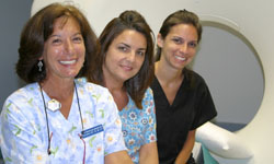
 We have a female staff specially trained in women’s health issues. Our technologists are highly trained and experienced. In addition to accuracy, they are devoted to your comfort before, during and after your tests are completed. We have a female staff specially trained in women’s health issues. Our technologists are highly trained and experienced. In addition to accuracy, they are devoted to your comfort before, during and after your tests are completed.
Karen Cortellino, M.D., and Dr. Charles Whang will review the study results with the patient on the same day of testing. She will answer any questions you may have. She will send the results to your doctor and the patient receives a report in the mail.
The Women’s Radiology Services that we offer are listed here and detailed below.
For information on how to prepare for your test, click here to view our Test Preparation Guide.
Maternity and Prenatal Services
High Mountain Health has been helping expectant mothers prepare for the healthy birth of their children for years. We offer a range of prenatal testing, the most common being the Ultrasound.
An Ultrasound consists of a series of high-frequency sound waves to form a photographic image of the developing fetus at different stages of the pregnancy.
Obstetrical/Fetal Ultrasound scans the uterus, ovaries and the fetus to determine healthy development and fetus size appropriate for its age.
As with other ultrasounds, a gel is applied to the area being scanned and a wand, or “transducer” is moved over the surface. This produces a sonographic, digitized image that details the fetus’ development.
Pregnancy Ultrasounds are repeated during the second and third trimester of the pregnancy. In preparation, the patient drinks three glasses of water one hour prior to the exam and does not void until the exam is completed.
The ultrasounds described above generally take 30 minutes.
Breast Imaging Services
 High Mountain Health is an accredited mammography facility by the American College of Radiology and performs more than 3,500 mammography procedures each year. High Mountain Health is an accredited mammography facility by the American College of Radiology and performs more than 3,500 mammography procedures each year.
Dr. Cortellino and Dr. Whang interpret standard mammographies as well as breast ultrasound examinations and breast MRIs, as may be requested by your doctor to provide the most accurate diagnosis.
What is a mammogram?
A mammogram is a safe, effective, low-dose X-ray that evaluates breast tissue. A screening mammogram can detect extremely small breast cancers that are too small to be discovered in a self-examination, providing them benefit of early detection, which is so important in effectively treating breast cancer in its earliest stage.
Diagnostic mammograms take additional X-rays for further study and are prescribed by your physician or the radiologist reviewing your mammogram.
What should I expect?
The mammogram is a simple procedure that generally takes no more than 20 minutes to perform. It should be scheduled when the breast is least tender, usually within the first ten days of the menstrual cycle.
When you arrive at High Mountain Health’s Radiology and Women’s Health office you will be greeted by an experienced staff member who will register you. Please bring your insurance card, prescription and previous mammograms.
A radiology technologist will situate you on the X-ray console and then take X-rays of each breast from above and from each side.
A Screening Mammography is an X-ray examination of the breasts where no symptom is apparent and there is no family history of breast cancer. The purpose of this test is to detect any sign of breast cancer that is too small to be detected by self examination. The goal is early detection, since this improves the opportunity for successful treatment. A screening mammography is recommended every year for women over 40.
A Diagnostic Mammography is an X-ray of the breast in a woman who has detected a symptom, such as a lump. This test is used to pinpoint the precise location of the abnormality as well as to image the surrounding tissue and lymph nodes. Further same-day testing may include a breast ultrasound or MRI.
A Breast MRI or Magnetic Resonance Mammography is used to obtain a more comprehensive view of the breast tissue and to determine the development of breast cancer disease. It may be necessary to schedule a Breast MRI at a later date if the woman is on hormone replacement, or is in the first or fourth week of her menstrual cycle.
A Breast MRI requires an injection of Gadolinium, which is a contrast material that increases the visibility of body tissue in the MRI images.
The American Cancer Society recommends an annual Breast MRI form women who are at “an extraordinarily high risk” of developing breast cancer in their lifetime. The ACS defines these risk candidates as:
- Women who carry the two breast cancer mutations – BRCA1 and BRCA2 genes, which are easily testable.
- Women who are first-degree relatives of someone with one of those gene abnormalities but have not yet been tested.
- Or a woman with a significant family history, who by a breast-cancer risk model, would have a lifetime risk of somewhere between 20 and 25 percent.
A Breast Ultrasound is used to determine whether a breast mass that has been identified is filled with fluid or is solid. It is not a substitute for standard mammographic exams.
Bone Scan, or Bone Densitometry
These tests measure mineral content in the bones. It involves an extremely small dose of radiation that determines bone mineral density (BMD). It compares your measurements to a reference population based on age, weight, sex and ethnic background. Physicians use this information to diagnose bone status and fracture risk. Low bone density is caused by osteoporosis, causing bones to become brittle. If detected, preventive therapy can be prescribed to slow or halt bone loss and, in some cases, reverse it.
|

 We have a female staff specially trained in women’s health issues. Our technologists are highly trained and experienced. In addition to accuracy, they are devoted to your comfort before, during and after your tests are completed.
 High Mountain Health is an accredited mammography facility by the American College of Radiology and performs more than 3,500 mammography procedures each year.